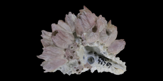
Still talking about animal fossils—this time, we’re diving into one of SpongeBob’s friends. Can you guess who it is? That’s right! One of our main topics today is Patrick, who is actually an example of an echinoderm. In addition to echinoderms, I’ll also be covering the arthropod group. Let’s check them out one by one!
ECHINODERMATA
A. Characteristics
- Their bodies are spiny.
- Triploblastic.
- Exoskeleton made of calcium carbonate (CaCO₃).
- High regenerative ability.
- Bilateral symmetry in the larval stage.
- Pentaradial symmetry in adulthood.
B. Taxonomix Level
- Phylum: Echinodermata
C. Classification
a. Asteroidea (sea stars)
- Star-shaped with ambulacral grooves.
- Have five arms/tentacles.
- Fossils are rarely found.
 |
| Aboral surface |
 |
| Oral surface |
b. Ophiuroidea (brittle stars)
- Do not have ambulacral grooves.
- Have long, slender, and highly flexible arms.
- Star-shaped body.
- Rarely found as fossils because their skeletons disintegrate quickly after death. When fossils are found, they are often well-preserved due to rapid burial and fossilization.
 |
| Aboral surface |
 |
| Oral surface |
c. Echinoidea (sea urchins)
-
Shaped like an egg, heart, or globular form.
-
Have many plates on their body.
-
Ambulacral grooves are perforated with pores.
-
Commonly found as fossils.
-
There are two types of echinoidea:
Regular: clearly displays pentameral symmetry.
-
Irregular: pentameral symmetry is not clearly visible.
 |
| Regular echinoidea |
d. Holothuroidea (sea cucumbers)
- Have soft, fine spines.
- Move flexibly but slowly.
- Mouth located at the anterior end and anus on the aboral side.
e. Crinoidea (sea lilies)
- Arms are segmented.
- Body shaped like a cup.
- Mouth and anus are located on the upper surface.
f. Blastoidea (extinct)
- Similar to crinoidea, but differs in the theca.
- The body has 5 prominent radiating grooves.
- There are 5 V-shaped plates.
g. Eocrinoidea (extinct)
- Similar to crinoidea, but differs in the theca.
h. Edrioasteroidea (extinct)
- Small fossils grow on top of edrioasteroids.
- Resembles a starfish.
- The organism is round with 5 tentacles.
- The tentacles grow in a curved, spiral, or near-spiral pattern.
i. Helicoplacoidea (extinct)
- Does not have pentameral vascular symmetry.
- The skin is covered with overlapping spiral ossicles that function like armor.
j. Arkarua (extinct)
- Shaped like a small disc.
- Resembles an echinoderm but in a smaller size.
D. Morphology
 |
| Asteroidea |
F. Index Fossils
- Helicoplacoidea: Cambrian
- Eocrinoidea: Cambrian–Silurian
- Edrioasteroidea: Ordovician
- Psammechinus: Pliocene
- Hemicidaris: Jurassic
- Acrosalenia: Jurassic–Cretaceous
- Coelopleurus: Eocene
- Clypeaster: Eocene
- Micraster: Cretaceous–Paleocene
- Holaster: Cretaceous
ARTHROPODA
 |
| Life form |
 |
| Fossil form digitalatlasofancientlife.org |
A. Characteristics
- The body is covered by a waxy substance/cuticle.
- The exoskeleton is made of protein/chitin.
- Coelomates.
- Legs are jointed.
- The body has bilateral symmetry.
B. Taxonomix Level
- Phylum: Arthropoda
C. Classifications
a. Crustacea (shrimp-like animals)- Has 4 antennae on the head.
- Has thoracic legs.
- The head and thorax are covered by a carapace.
- Lives in freshwater, marine, and terrestrial environments.
- Some examples of crustacea:
 |
| Fossil form digitalatlasofancientlife.org |
 | ||
Life form
|
b. Chelicerata
- Has specialized mouthparts called chelicerae that allow early members of the group to feed on others.
- Chelicerata consists of the following classes:
 |
| Life form |
 |
| Fossil form digitalatlasofancientlife.org |
 |
| Fossil form digitalatlasofancientlife.org |
 | ||
Life form
|
 |
| Fossil form digitalatlasofancientlife.org |
d. Myriapoda (centipedes)
- Consists of only the head and body.
- There is one pair of eyes, sensory organs, and mouth on the head.
- Each segment has jointed legs.
E. Trilobite
Actually, trilobites have their own subphylum called Trilobitomorpha. However, I intentionally separated the class Trilobita as a main discussion topic because trilobites have very diverse shapes, making them useful as indicators of certain regions and specific ages. Additionally, there are already maps showing the distribution of trilobite habitats, commonly known as trilobite provinces.
These different forms are due to:
-
Evolution progressing from simple to more complex shapes.
-
Their habitat, such as the availability of food sources, which can affect the size of the trilobite.
Trilobites lived around the Cambrian to Permian periods. They became extinct during the Permian period due to extreme drying of the Earth's climate. Generally, trilobites can be divided into three body parts that are key for identification:
-
Cephalon
-
Thorax
-
Pygidium
 |
| Morphology of trilobites |
A. Trilobites can be distinguished based on:
a. The size ratio between the cephalon and the pygidium
- Micropygous: cephalon size is much larger than the pygidium. Example: Redlichia.
- Subisopygous: cephalon and pygidium sizes are nearly equal. Example: Bathyurus.
- Isopygous: cephalon and pygidium are the same size. Example: Ogygiocaris.
- Macropygous: pygidium size is larger than the cephalon. Example: Scutellum.
The general evolution of trilobites can be seen in the image below:
It can be concluded that trilobite morphology evolved starting from an elongated form. In the early stages, their morphology was very complex. Over time, the segments in the thorax began to reduce and their eyes started to disappear. Then spines began to appear while other parts continued to diminish. This reduction process continued until trilobites became extinct in the Permian period.
In fact, there are several processes that accompany trilobite evolution, including:
1. Effacement
Some trilobite orders, especially Agnostida, Corynexochida, and Asaphida, experienced reduction or simplification in parts of the cephalon, thorax, and pygidium. This is related to the trilobites’ adaptation to a burrowing lifestyle. The effacement can be seen in the image below:
Spinocity can be defined as the growth of spines on trilobites, which can arise from almost any part of the exoskeleton, especially along the margins. For example, the extension of the pleura (segments on the thorax). The growth of these spines is a defensive adaptation to protect themselves from predators, indicating that life during the Paleozoic era was very dangerous for trilobites.
Miniaturization is the reduction in size of trilobites, usually caused by a decreasing food supply. This can happen for two reasons: the trilobite population increases, leading to higher competition for food, or the food itself becomes scarce. The purpose of reducing their body size is so that trilobites require less food to survive, helping to maintain the available food resources.
C. Some Trilobite Morphologies
1. Atheloptic Morphology
Trilobites living in deep marine environments are characterized by absent or reduced eyes, as an adaptation to their deep, low-light, aphotic habitat.
2. Pelagic Morphology
They have large eyes and an elongated, slender body shape as adaptations to enable trilobites to swim well and disperse across the entire ocean (long-distance travel).
3. Olenimorph
The exoskeleton is thin, the number of thorax segments increases, and the body shape is flat and wide as an adaptation to low oxygen levels and high sulfur concentrations. The various transversal thorax pleurae cover the gills and extend laterally, allowing for maximum oxygen absorption and providing a large surface area for symbiotic bacteria (sulfur-eating bacteria as a feeding strategy) to live.
4. Pitted Fringe
The expansion of the cephalon into a concave space is an adaptation for filter feeding (helping to stabilize the trilobite during filtration).
D. Habitat
E. Types of Trilobites based on Geological Time Scale


0 comments:
Post a Comment